CTmeasure (integrated CT measurement software)
CTmeasure (current version: 0.96a)
The official integrated software for CT image measurements provided by Japanese Society of Computed tomography Technology (JSCT)
- image viewing window
- Analysis window
1. Introduction
CTmeasure has been developed to facilitate physical CT image measurements such as modulation transfer function (MTF), noise power spectrum (NPS), section sensitivity profile (SSP), etc.
This software is one of membership contents provided by 'CT measure production board' (Director: Katsuhiro Ichikawa, Ph.D), which is one of several special boards in JSCT. Therefore, you should be a member for using this software, and naturally anyone who is not a member can not use it. So far, foreigners can not use this software because there is no English web page for the membership. We are planning to make a English web page for providing CTmeasure at a specific price and/or to make a free version of CTmeasure with limited functions.
2. Concept
CTmeasure consists of two windows: Image viewing window and Analysis window. A target image or a target image range (start and end images) can be set, viewing images on Image viewing window using paging style. After the target image (range) selection, the user analyzes the image (range) by selecting one of functions in Analysis window. The target image range selection is very important to improve the accuracy of MTF, NPS, CT-value, and SD, giving users averaged results over the image range, and very efficient to obtain the accurate results shortly without repeated operations. Of course, the image range selection is essential for SSP and temporal sensitivity profile (TSP) which naturally need multiple images.
This software has been authorized by the board members, and in addition been improved based on the users' evaluation (feed backs). Therefore, CT measure production board believes that this software would contribute to various studies on CT technology including CT image measurements.
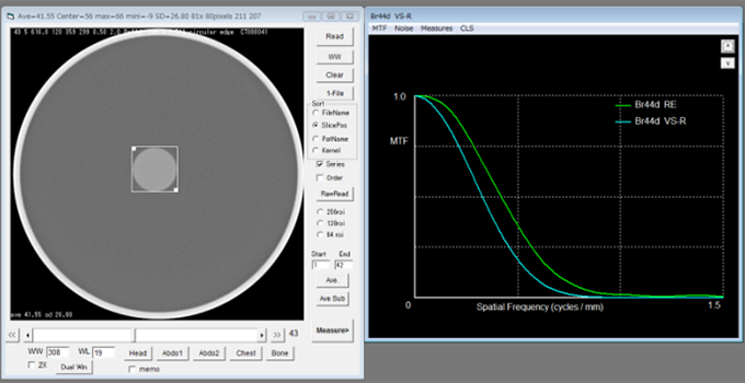
Screen shot in MTF analysis using the circular edge method
3. Available measurements
CTmeasure provides functions below. All of algorithms are based on the previously published papers listed in Reference section.
1) Image viewing window
CT value and standard deviation (SD) measurements, preset windows, 2X magnify, image averaging and subtraction, dual window viewing (comparison mode), and raw format reading are available. Unlike the famous image analysis software, image J, the window width and window level can be adjusted by mouse drugging similar to clinical CT systems. The CT and SD values are always displayed as overlays, which provides an easy operation (non-operation) for such measurements. The user can set the start and end image numbers for analysis using multiple images by simply clicking at 'Start' and 'End' text boxes.
2) Analysis window
a) MTF analysis
MTF can be analyzed from wire images and the task-based MTF (MTFTASK) for iterative reconstructions (IRs) can be also calculated from disk images and linear edge images.
The 2-dimensional (2D) fast Fourier transformation (2D-FFT) method [1-3], the virtual slit (synthesized slit) method and the radial virtual slit method for wire images are available.
The virtual slit method has been used for NPS analysis of digital radiography to obtain a central slice of the 2D NPS [4, 5]. By using the same concept, a 1D line spread function (LSF) can be obtained from point spread function (PSF) images which can be obtained by scanning a thin metal wire phantom (the wire method: conventional MTF measurement method for CT). In the radial virtual slit method, 18 LSFs, which are obtained by rotating the scanning direction of the virtual slit method with intervals of 20 degree, are averaged, and thereby more smoothed (accurate) LSF can be obtained.
In the (radial) virtual slit method, it is easy to visually inspect the LSF (i.e. 1D profile of wire image) and to eliminate the unnecessary regions distant from the LSF center (making the regions zero). The agreement between the virtual slit method in CTmeasure and the 2D FFT method was confirmed in reference #6 and by repeated validation tests by the board members.
The circular edge method for disk images and linear edge method for edge images obtained by scanning a flat surface in a block phantom are also available. These methods can be used for recent IR images by using CT images with different contrasts and noise levels, based on the task based consideration [7].
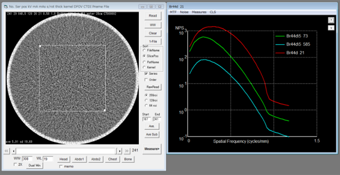
b) NPS analysis
Radial frequency method using the 2-dimensional (2D) fast Fourier transformation (FFT) [4,5,8] and virtual slit method using the 1D FFT [1-3] are available. In advance of analysis, one of three ROI sizes of 64x64, 128x128, and 256x256 should be selected in Image viewing window. The frequency bin technique for obtaining smoothed NPS data presented in an IEC standard [9] is utilized. The frequency bin width is selectable in 0.02/pixel pitch (data number = 24) or 0.01/pixel pitch (data number = 48). Though the frequency bin technique was recommended for obtaining smoothed NPS curves for digital radiography, it is also effective for CT NPS analysis [10].
c) z-directional resolution analysis(slice thickness)
z-directional (cross section) spatial resolution can be evaluated by SSP. A thin metal disk and a small metal bead have been recommended to approximate the delta signal [11-13]. Since thin slice evaluations are frequently needed for recent CT systems, the thin metal disk and the bead need considerations about disk alignment error and low response of too small bead, respectively. To reduce these drawbacks, a micro coin (1-mm diameter and 0.05-mm thickness) method was reported [14,15], and the CT measure production board also recommends this micro coin method. Full width at half maximum (FWHM) and tenth maximum (FWTM) results can be easily obtained using CTmeasure and in addition the z-directional MTF can be analyzed from the SSP profile.
d) Temporal resolution analysis
The temporal sensitivity profile (TSP) can be measured by scanning a metal ball passing through the scan plane in the direction along z-axis at a high speed [16, 17]. This measurement enables to know actual temporal resolution of not only the helical scan mode but also the cardiac mode without multi-segments. The FWHM and FWTM analysis and the temporal transfer function (temporal TF) calculation are also available.
e) CT profile analysis
A CT number profile can be obtained from a rectangular ROI placed on images in a target range.
4. Feature plan of CTmeasure
CTmeasure is not free software, and thus foreigners are difficult to get this software because the membership web page in English does not exist now. To allow the foreigner to use this software, we are planning to open the English web page to get only CTmeasure at a cheaper price than the membership price (about 30 US dollar for a membership), or to develop a free version of CTmeasure (e.g. CTmeasure basic) with limited functions.
Reference
[1] Bischof CJ and Ehrhardt JC : Modulation transfer function of the EMI CT head scanner. Med. Phys. 4(2), 163-167, (1977)
[2] Boedeker KL, Cooper VN, McNitt-Gray MF. Application of the noise power spectrum in modern diagnostic MDCT: part I. Measurement of noise power spectra and noise equivalent quanta. Phys Med Biol. 2007 52(14):4027-4046.
[3] Ichikawa K, Kobayashi T, Sagawa M, Katagiri A, Uno Y, Nishioka R, Matsuyama J, A phantom study investigating the relationship between ground-glass opacity visibility and physical detectability index in low-dose chest computed tomography, J Appl Clin Med Phys. 16(4): 202-215, 2015
[4] M. L. Giger, K. Doi, and C. E. Metz, ‘‘Investigation of basic imaging properties in digital radiography. 2. Noise Wiener spectrum,’’ Med. Phys. 11, 797-805 ~1984.
[5] Siewerdsen JH, Antonuk LE El-Mohri Y, et al.: Signal, noise power spectrum, and detective quantum efficiency of indirect-detection flat-panel imagers for diagnostic radiology, Med Phys, 25(5), 614-628, (1998).
[6] Ichikawa K, Hara T, Niwa S, Ohashi K, Method of measuring modulation transfer function using metal wire in computed tomography. Nihon Hoshasen Gijutsu Gakkai Zasshi. 2008, 64(6): 672-80 (in Japanese)
[7] Richard S, Husarik DB, Yadava G, Murphy SN, Samei E.Towards task-based assessment of CT performance: system and object MTF across different reconstruction algorithms. Med Phys. 2012 ;39(7):4115-4122.
[8] Marie Foley Kijewski and Philip F Judy, The noise power spectrum of CT images Phys. Med. Biol., 1987, Vol. 32, No 5, 565-575.
[9] IEC62220-1:Medical electrical equipment-Characteristics of digital X-ray imaging devices part1:Determination of detective quantum efficiency.(2003).
[10] Ichikawa K, Hara T, Niwa S, Yamaguchi I, Ohashi K, Calculation Methods for Noise Power Spectrum Measurement in Computed Tomography, Medical Imaging and Information Sciences 2008; 25(2), 29-34 (in japanese)
[11] Polacin A, Kalender WA, and Marchal G: Evaluation of section sensitivity profiles and image noise in spiral CT, Radiology, 1992; 185, 29-35.
[12] Polacin A, Kalender WA, and Brink J, et al.: Measurement of slice sensitivity profiles in spiral CT. Med Phys, 1994; 21(1), 133-140.
[13] Slice-sensitivity profile and noise. In: Jiang Hsieh. Computed tomography: principles design artifacts and recent advances. Bellingham, WA: SPIE Press; 2009. 348-354.
[14] Hara T, Tsuzaka M, Sakurai N. Measurement of modulation transfer function in Z-axis for multi-slice spiral CT using the micro-disk method: comparison with the bead method and examination of geometric influence, Nihon Hoshasen Gijutsu Gakkai Zasshi. 2003;59(11):1391-8. (in Japanese)
[15] Urikura A, Nakaya Y, Ichikawa K, Kawatani K, Kawashima I, Goto H, Physics properties of non-helical scan using 320-row multi detector computed tomography,.Nihon Hoshasen Gijutsu Gakkai Zasshi. 2012;68(4):432-42. (in japanese)
[16] Ichikawa K, Takada T, Hara T, Ohashi K, Niwa S, A new method of measuring temporal resolution for computed tomography. Nihon Hoshasen Gijutsu Gakkai Zasshi. 2008; 64(9):1172-1176. (short report in Japanese)
[17] Ichikawa K, Hara T, Urikura A, Takata T, Ohashi K, Assessment of temporal resolution of multi-detector row computed tomography in helical acquisition mode using the impulse method. Phys Med. 2015;31(4):374-381.
Board Members
- Katsuhiro Ichikawa, Ph.D.
- Institute of Medical, Pharmaceutical and Health Sciences, Kanazawa University
- Takanori Hara Ph.D.
- Department of Medical Technology, Nakatsugawa Municipal General Hospital
- Kazuya Ohashi, Ph.D.
- Department of Central radiology, Nagoya University
- Kazuhiro Satoh, Ph.D.
- Tohoku University Hospital